Multiple Sclerosis Mri Brain White Matter Lesions

New Ms Subtype Shows Absence Of Cerebral White Matter

Imaging In Multiple Sclerosis Journal Of Neurology Neurosurgery

The Multiple Sclerosis Lesion Checklist Practical Neurology
Brain Imaging In Multiple Sclerosis Practice Essentials Computed
:max_bytes(150000):strip_icc()/what-are-these-spots-on-my-mri-2488902-5c5db0fa46e0fb0001ca86cb.png)
Spots On An Mri White Matter Hyperintensities
Https Encrypted Tbn0 Gstatic Com Images Q Tbn 3aand9gcr1ryuhls8ao8r6aab6o8r32bwv7kljtuinbg Usqp Cau
You can see them in other diseases which involve the blood vessels of the brain causing vasculitis that is inflammation of the blood vessels and this list includes neurosarcoidosis.
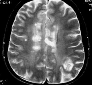
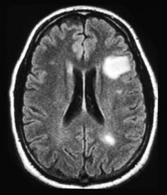
Multiple sclerosis mri brain white matter lesions. Specifically the periventricular lesions and the more peripheral white matter lesions near the gray matter white matter junction are typical mri findings in multiple sclerosis. There are several causes of white spots on a brain mri including small strokes migraines multiple sclerosis ms lupus b12 deficiency a brain tumor such as lymphoma or an infection such as lyme disease or hiv. Ms related lesions appear on mri images as either bright or dark spots depending on the type. This contention is based on the premise that cerebral demyelination signs on mri are sufficiently recognizable and characteristic to be considered a sine qua non of ms diagnosis.
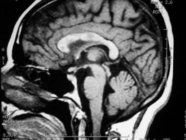
The role of mri in diagnosing ms if you have symptoms of ms your doctor may order an mri scan of your brain and spinal cord. The key question then is whether a patient s mri shows ms like lesions. White matter appears white because the protective wrapping around nerve fibers or axons is a pale fatty tissue called myelin. The images produced allow doctors to see lesions in your cns.
2 a corollary is that presence of multiple white matter lesions does not increase likelihood of ms as long as none or very few of the lesions are typical of ms. Axons are like the electric wires of the brain says rhonda. Mri scans can detect damage in the central nervous system which comprises the brain and spinal cord. 1 they are usually found in the brain s white matter typically near the ventricles but they can be located anywhere in the brain.

The Radiology Assistant Multiple Sclerosis

Periventricular White Matter Lesions

Differentiating Multiple Sclerosis Mimics On Mri Neurology Advisor

Periventricular White Matter Lesions
Multiple Sclerosis Practice Essentials Background Pathophysiology

Proton Density Weighted Images From Two Subjects With Multiple

Mri In Multiple Sclerosis A Hyperintense Parietal White Matter

Mri Features Of Benign Multiple Sclerosis Neurology

New Page 1

Differential Diagnosis Of Multiple Sclerosis And Other

White Matter Lesions Diagnostic Image Analysis Group
Brain Imaging In Multiple Sclerosis Practice Essentials Computed

Periventricular White Matter Lesions
