Ovarian Cancer Ultrasound
Malignant Ovarian Tumor Imaging Practice Essentials Computed
Imaging Techniques For The Diagnosis Of Ovarian Cancers

A Better Way To Assess Ovarian Cancer Risk Ncal Research Spotlight
Ovarian Tumors
What 3d Ultrasound Can Tell You About Ovarian Cancer Empowered

Iota Simple Rules And Srrisk Calculator To Diagnose Ovarian Cancer
It can also be used to get a better look at the ovary to see how big it is and how it looks inside.
Ovarian cancer ultrasound. Ultrasound is often the first test done if a problem with the ovaries is suspected. Many women with ovarian cancer have. The ca 125 blood test measures the amount of a protein called ca 125 in the blood. A positron emission tomography pet scan isn t usually used to check for ovarian cancer.
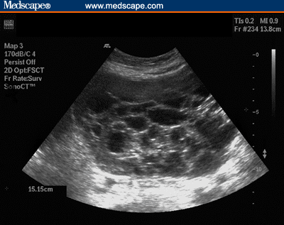
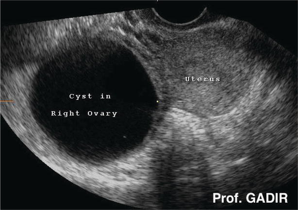
Ultrasound scans use high frequency sound waves to create a picture of a part of the body. During an external ultrasound of your pelvis the doctor or radiographer moves a probe over the lower part of your tummy. Transvaginal ultrasound or tvus usually prescribed for screening and detecting ovarian cancer uses sound waves for looking inside the uterus ovaries and fallopian tubes. You might have an external ultrasound of your lower tummy pelvis or a vaginal ultrasound to help diagnose ovarian cancer.
Pet uses radioactive glucose to spot. By the time the changes of ovarian cancer are detectable by ultrasound most ovarian cancers are well beyond the early stage of the disease. This technology uses sound waves to create an image of internal organs including the ovaries uterus and cervix. It can show the ovaries womb and surrounding structures.
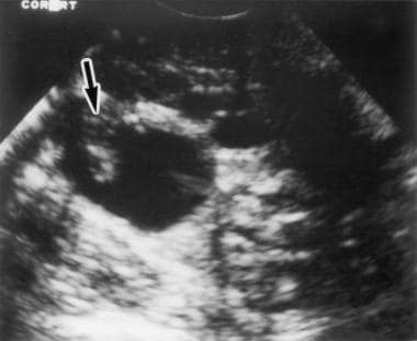
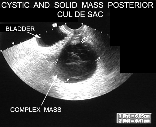
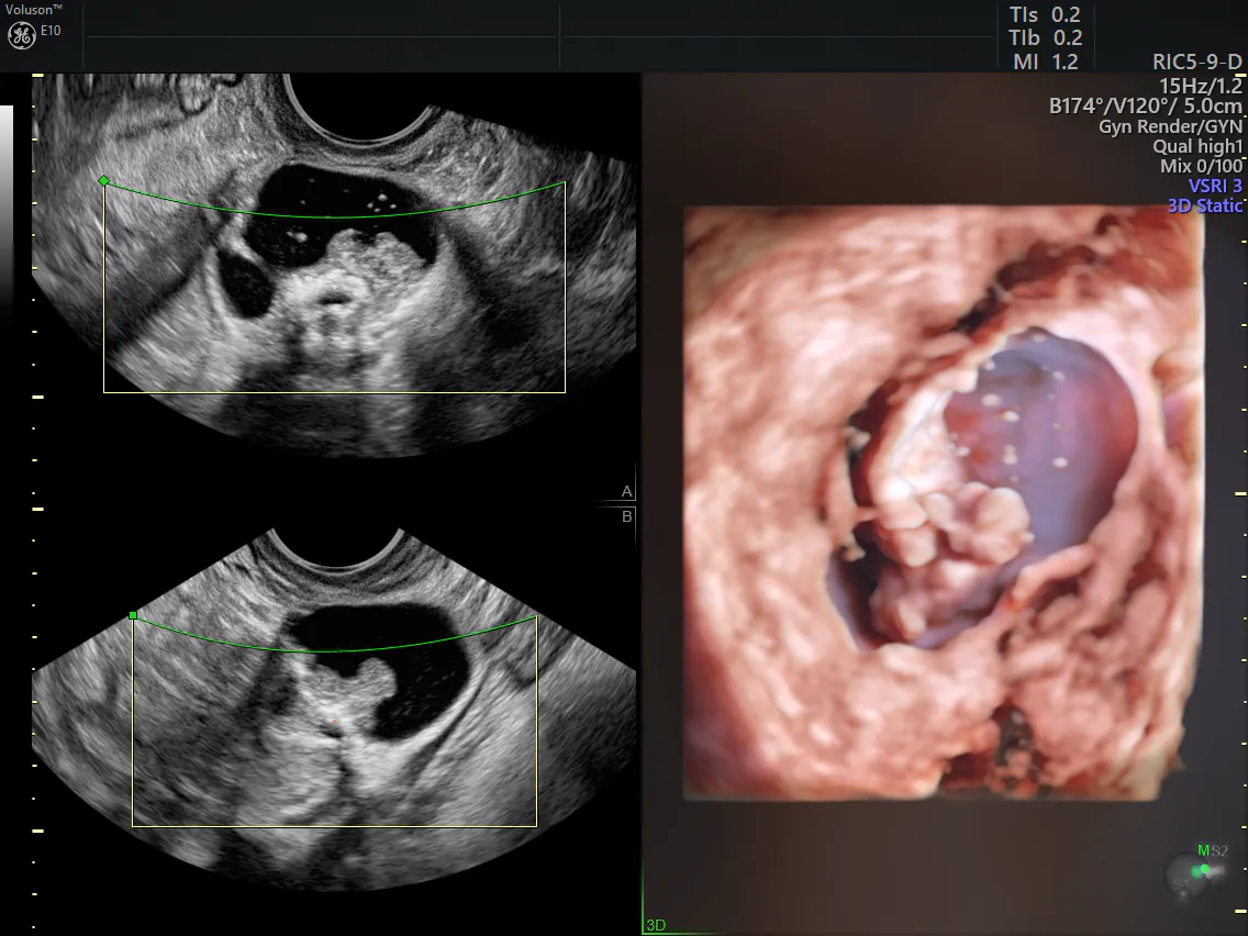
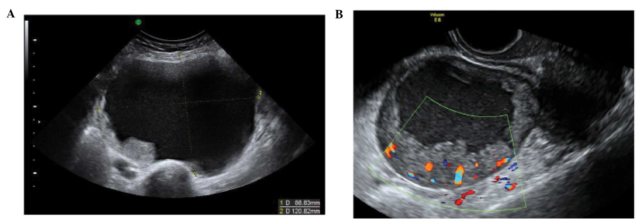
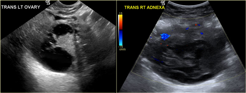
It s more useful in finding out whether ovarian cancer has spread. The process of tvus uses an ultrasound wand into the vagina in order to find any mass or lump in the ovary or uterus though not all the masses found would essentially be tumor. The sound waves that bounce off cancerous tissue are different than those reverberating off healthy tissue allowing an ultrasound to differentiate between normal tissue and some tumors.
Ultrasound Imaging Of Ovarian Cancer Chapter 22
Ovarian Cancer And Pregnancy Intechopen
Diagnostics Free Full Text Ultrasound Monitoring Of Extant

Pelvic Ultrasound In Houston For Ovarian Cancer Screening
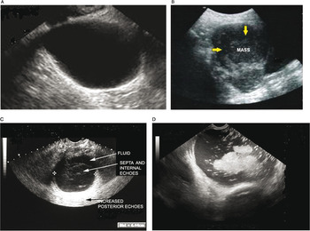
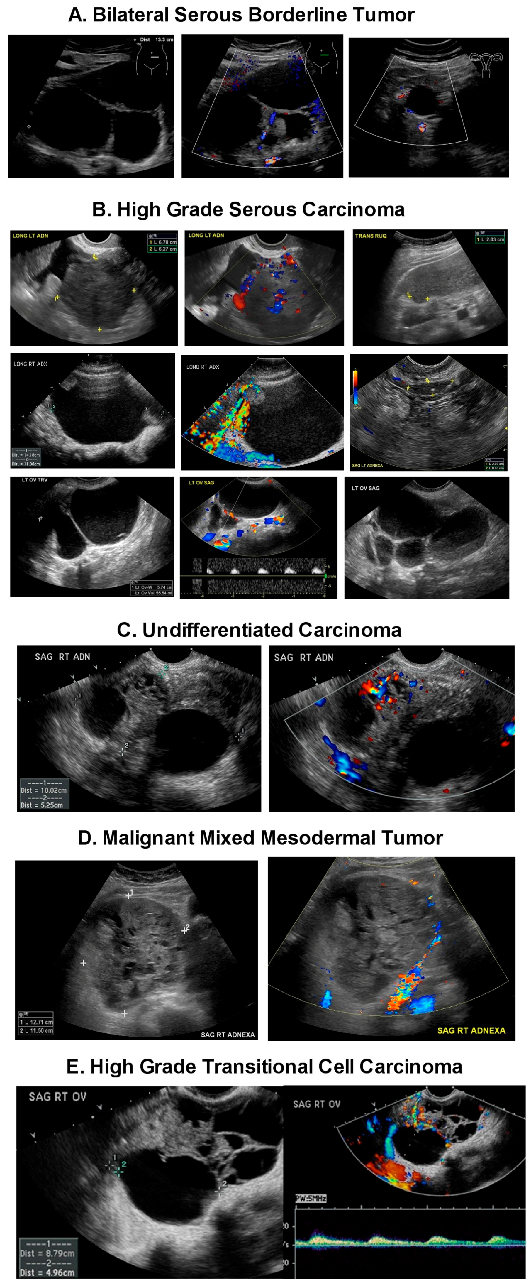
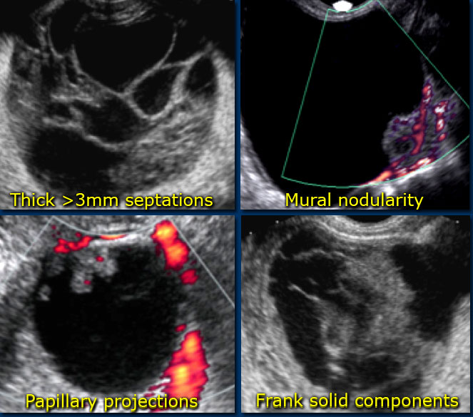
The Characteristic Ultrasound Features Of Specific Types Of

Figure 1 From Arly Detection Of Ovarian Cancer By Tumor Epithelium

Usf Health News Watchful Waiting With Routine Ultrasound Safer
Ovarian Tumors

The Enhancement In Ultrasound Signal Intensity From Spontaneous

Ultrasound Based Screening For Ovarian Cancer Leaders In
Detection And Diagnosis Ovarian Cancer Symptoms And Diagnosis
The Radiology Assistant Roadmap To Evaluate Ovarian Cysts
The Radiology Assistant Common Ovarian Cystic Lesions
