Mri Findings In Parkinson S Disease Radiographics
Clinical Application Of Brain Mri In The Diagnostic Work Up Of

The Substantia Nigra In Parkinson Disease Proton Density Weighted
Parkinson Disease Radiology Reference Article Radiopaedia Org

Imaging In Parkinson S Disease Practical Neurology
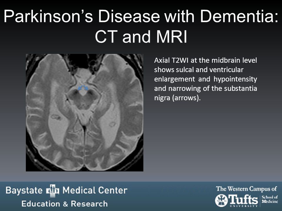
Presentation1 Pptx Radiological Imaging Of Parkinsonism
Structural Mri In Idiopathic Parkinson Disease And Parkinsonism

Classically 5 hz pill rolling resting tremor.
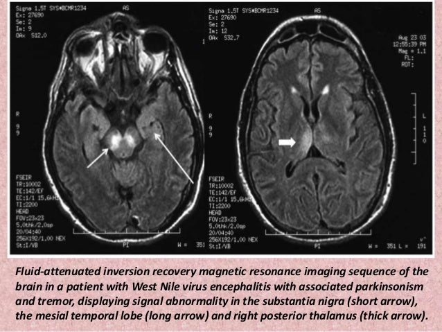
Mri findings in parkinson s disease radiographics. Although these cannot be definitively diagnosed or differentiated from parkinson s disease pd with mri there are radiologic hallmarks that can be useful when combined with the clinical examination and other diagnostic tests. Magnetic resonance parkinsonism index maung maung soe and dr marcin czarniecki et al. Toxic and metabolic brain disorders manifest secondary to derangements of a well balanced environment encompassing metabolic substrates neurotransmitters electrolytes physiologic ph levels and blood flow either by endogenous malfunctions or exogenous toxic effects. This article addresses some of the most challenging diagnostic issues in neuroimaging.
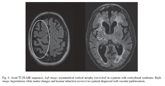
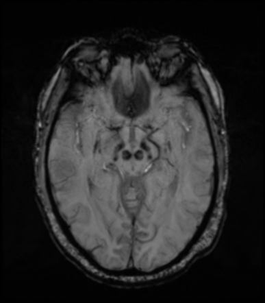
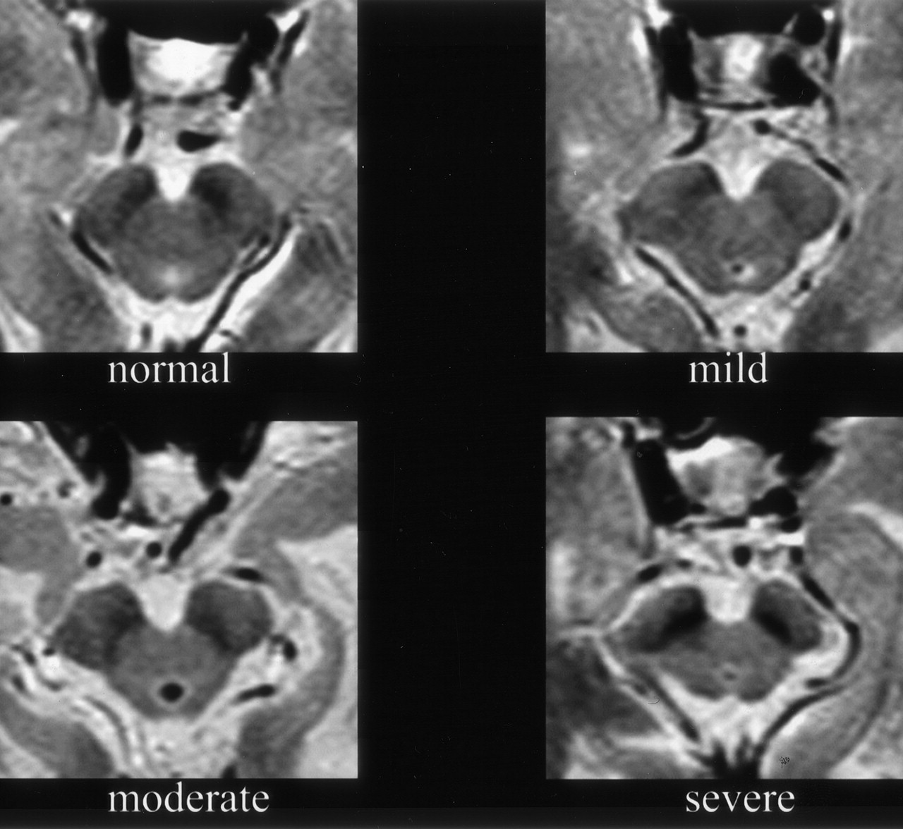
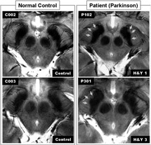
However the primary use of mr imaging is to exclude specific structural abnormalities that could potentially mimic parkinson disease eg normal pressure. Share on pinterest an mri or ct scan can help to distinguish parkinson s from other conditions that may have similar symptoms such as a stroke. Fdg pet uptake can demonstrate patterns of neuronal dysfunction that are specific to a particular parkinsonian syndrome. In advanced disease abnormalities of the substantia nigra including volume loss decreased t2 signal reflecting iron deposition and blurring of the margins can be seen 26 28.
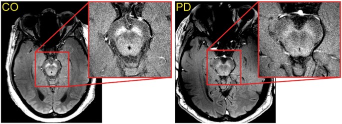
The classic cardinal motor features of parkinson disease which are often asymmetric include. Although no single imaging test is diagnostic a combination of tests may help narrow the differential diagnosis. Our purpose was to determine the exact location of the substantia nigra by correlating imaging and. The discovery which has the potential to revolutionise the diagnosis of this important disease uses axial high resolution susceptibility weighted imaging swi to assess the structure of the substantia nigra within the midbrain.
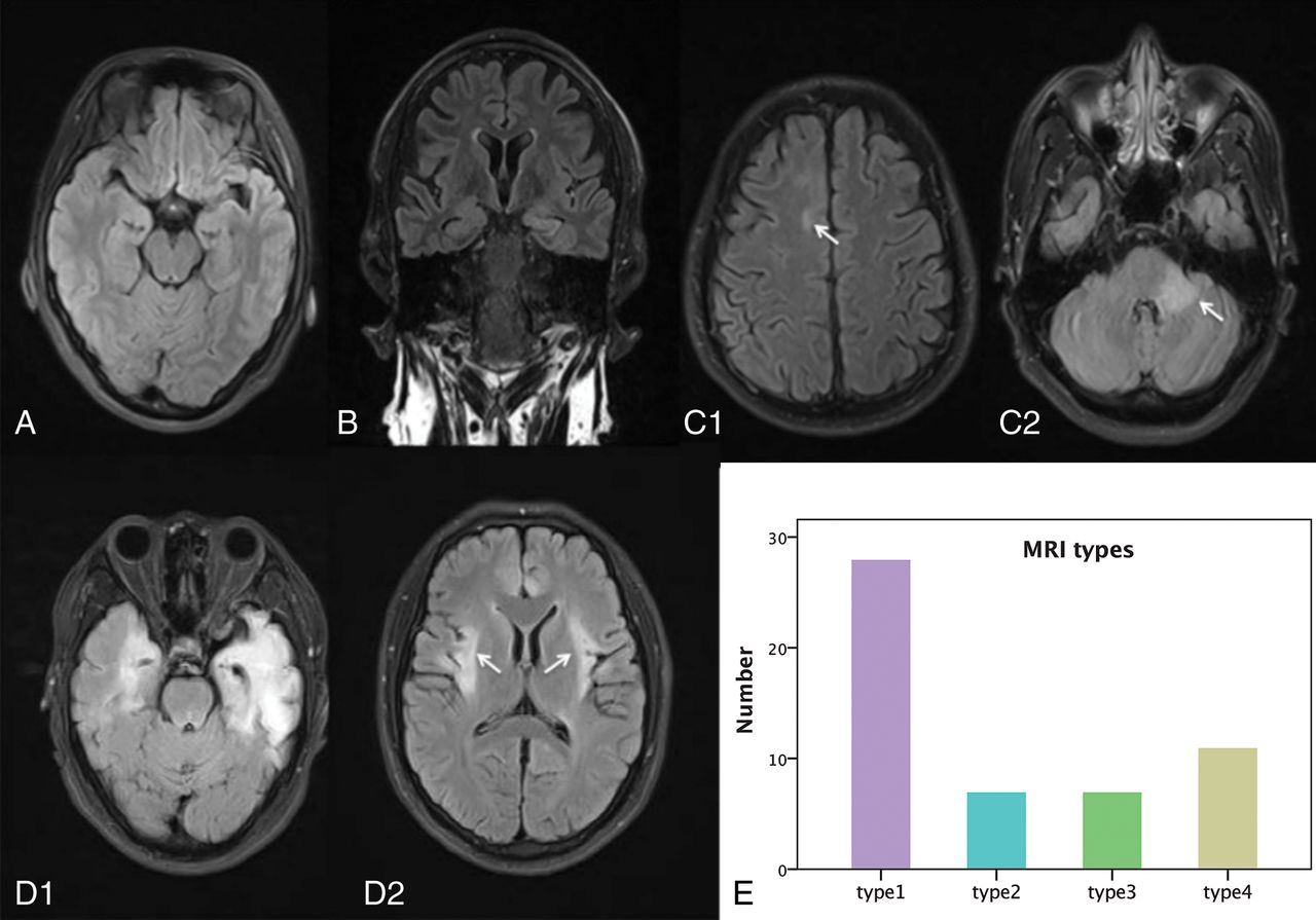
Most prominent in the distal upper limbs. Unlike parkinson disease and lewy body dementia two other synucleinopathies these intracellular deposits are found not only in neurons but also in oligodendroglia 2. Mri signs of parkinson s disease mimics. Conventional mr imaging is usually not helpful in the diagnosis of early parkinson disease because it most often yields normal findings.
Several neurodegenerative diseases can cause parkinsonian motor symptoms. A recent research publication in plos one has described a new 3t mri sign of parkinson disease known as the absent swallow tail sign. By dr andrew dixon. In 2016 experts developed new criteria for.
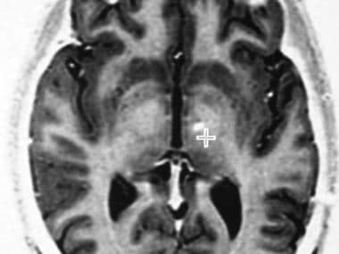
The substantia nigra is anteroinferolateral to the red nucleus and it is important to precisely locate its true anatomic location to accurately measure sn area. Findings at 123 i ioflupane spect can confirm the loss of dopaminergic neurons in patients with parkinsonism and help distinguish these syndromes from treatable conditions including essential tremor and drug induced parkinsonism. Magnetic resonance parkinsonism index mrpi can be used in mri studies to predict the presence of progressive supranuclear palsy psp in patients with clinically unclassifiable parkinsonism.

Mri In Parkinson Disease Expanding Usability For Better
Mr Imaging Of The Superior Profile Of The Midbrain Differential

Pdf Brain Mri In Parkinson S Disease Semantic Scholar
When Is Mri Indicated In The Evaluation Of Patients With Suspected
Nigrosome 1 Visibility At Susceptibility Weighted 7t Mri A
Unforgettable Images A Multimodality Pictorial Review Of Dementia
Brain Mri Characteristics Of Patients With Anti N Methyl D

Unilateral Absence Of Swallow Tail Sign In Parkinson Disease
Imaging And Behavior In Parkinson S Disease Structural Imaging

Loss Of Substantia Nigra Hyperintensity On 7 Tesla Mri Of
Neuromelanin Detection By Magnetic Resonance Imaging Mri And Its

Comparative Study Of Mri Biomarkers In The Substantia Nigra To
Fig 4 Nigrosome 1 Detection At 3t Mri For The Diagnosis Of
